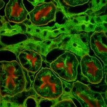
Principle:
Viable cells take up dye, diacetyl fluorescein and hydrolyze it to fluorescein to which the cell membrane of live cells is impermeable. Live cells fluoresce green and dead cells do not. Nonviable cells may be stained with propidium iodie and subsequently fluoresce red. Viability is expressed as the % of cells fluorescing green. This method can be applied to CCD or flow cytometry.
Outline:
Stain a cell suspension in a mixture of propidium iodide and diacetyl fluorescein and examine cells by fluorescence microscopy and flow cytometry
Materials:
Sterile or aseptically prepared:
Single cell suspension
Fluorescein diaacetate, 10ug/ml in HBSS
Propidium iodide,, 500ug/ml
Non-sterile:
Fluorescence microscope
Filters: Fluorescein : excitation 450/590 nm, emission LP 515 nm
Propidium iodide: excitation 488 nm, emission 615 nm
Protocol:
Prepare cell suspension as for dye exclusion but in medium without phenol red
Add fluorescent dye mixture at a proportion of 1:10 to give a final conc of 1ug/ml of 1ug/ml of diacetyl fluorescein and 50 ug/ml of propidium iodide
Incubate cells at 37oC for 10 min
Place a drop of cells on microscope slide, add coverslip and examine cells by fluorescence microscopy
B. Estimation of viability by dye exclusion
Principle:
Viable cells are impermeable to Napthalene Black, Trypan Blue and a number of other dyes
Outline:
Mix a cell suspension with stain and examine it by low power microscopy
Materials:
Sterile or aseptically prepared:
Cells for testing e.g. flask for trypsinization , frozen vial to thaw or primary disaggregate
Growth medium appropriate to cell type 20ml
Tryspin 0.25 % 5ml
D-PBSA 10ml
Non-sterile:
Hemocytometer
Viability stain (e.g. 0.4 % Trypan Blue or 1 % Napthalene Black in D-PBSA or HBSS)
Pasteur pipettes
Microscope
Tally Counter
Protocol:
Prepare a cell suspension at a high conc (~1 X 10⁶ cells/ml) by trypsinization or by centrifugation and resuspension
Take a clean hemocytometer slide and fix the coverslip in place
Mix one drop of cell suspension with one one drop (Trypan Blue) or four drops (Napthalene Black) of stain
Load the counting chamber of hemocytometer
Leave the slide for 1-2 min (do not leave any longer or viable cells will deteriorate and take up the stain)
Place the slide on microscope and use a 10X objective to look at the counting grid)
Count total number of cells and number of stained cells
Wash hemocytometer and return it to its box
Subscribe to:
Post Comments (Atom)

No comments:
Post a Comment